


Arthrosis of the shoulderIs chronic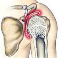 diseases in which articular cartilage tissue is destroyed and thinned, pathological changes in soft tissues occur, and bone growth is formed in the joint area. This is indicated by pain and cracking in the affected area. At a later stage, the range of motion decreases. The pathology is chronic and develops gradually. Diagnosis is made taking into account the clinical picture and radiological signs. Treatment is usually conservative: physiotherapy, anti-inflammatory drugs, chondroprotectors, exercise therapy. When the joint is destroyed, arthroplasty is performed.
General information
Arthrosis of the shoulder joint is a chronic disease in which, as a result of degenerative-dystrophic processes, cartilage and other joint tissues are gradually destroyed. Usually arthrosis strikes people aged 45 years and older, but in some cases (after injury, inflammation), the disease can develop at a younger age. The same pathology often occurs in women and men, more often observed in athletes and hard -working people.
The reasons
The starting point of change of arthrosis of the shoulder joint can be the process of normal aging of tissues and damage or disruption of cartilage structure due to mechanical influences and various pathological processes. Primary arthrosis is usually detected in the elderly, secondary (developing with a background of other diseases) can occur at any age. The main reasons considered:
- Developmental disabilities.Pathology can be detected with lack of the head of the humerus or glenoid cavity, shoulder capomelia and upper limb anomalies.
- Traumatic injuries.Traumatic etiological arthrosis most often occurs after intra-articular fractures. Possible causes of the disease are shoulder dislocations, especially the common ones. More rarely, severe bruising acts as a provoking injury.
- The inflammatory process.The disease can be diagnosed with long-term shoulder-scapular periarthritis, previously suffering from nonspecific purulent arthritis and special joint arthritis (with tuberculosis, syphilis and some other diseases).
Risk factors
Arthrosis is a polyetiological disease. There are a group of factors that increase the likelihood of this pathology:
- Hereditary tendencies.Many patients have close relatives who also suffer from arthrosis, including other localizations (gonarthrosis, coxarthrosis, arthrosis of the ankle joint).
- Excess joints.This can happen to volleyball players, tennis players, basketball players, sports equipment throwers, and even to people whose profession involves constant high loads on their hands (hammers, loaders).
- Other pathologies.Arthrosis is more frequently detected in patients suffering from autoimmune (rheumatoid arthritis), some endocrine diseases and metabolic disorders, lack of systemic connective tissue and excessive joint mobility.
The likelihood of this disease increases dramatically with age. Frequent hypothermia and adverse environmental conditions have certain negative effects.
Pathogenesis
The main reason for the development of arthrosis of the shoulder joint is a change in the structure of the articular cartilage. Cartilage loses its smoothness and elasticity, sliding of the articular surface during movement becomes difficult. Microtrauma occurs, which causes deterioration of the condition of cartilage tissue. Small cartilage pieces detach from the surface, forming an independent articular body, which also injures the inner surface of the joint.
Over time, the capsule and synovium thicken, areas of fibrous degeneration appear within them. Because of the thinning and decrease in elasticity, the cartilage stops providing the required shock absorption, therefore, the load on the underlying bone increases. The bone changes shape and grows along its edges. The normal configuration of the joint is disturbed, there are limitations of movement.
Welding
In traumatology and orthopedics, a three -stage systematization is commonly used, which reflects the severity of pathological changes and symptoms of arthrosis of the shoulder joint. This approach allows you to choose the optimal medical tactics, taking into account the severity of the process. The following levels are distinguished:
- The first- no major changes in cartilage tissue. The composition of synovial fluid changes, cartilage nutrition is disrupted. Cartilage does not tolerate stress, therefore, joint pain (arthralgia) occurs from time to time.
- The second- cartilage tissue begins to thin, its structure changes, the surface loses its smoothness, cysts and areas of calcification appear in the depth of the cartilage. The underlying bone is slightly deformed, bone growth appears along the edge of the articular platform. The pain becomes permanent.
- Third- Significant thinning and disruption of cartilage structure with extensive areas of destruction. Defective articular platform. Revealed movement limitations, ligament weakness and periarticular muscle atrophy.
Symptoms
In the early stages, patients with arthrosis are concerned about minor discomfort or pain in the shoulder joint while performing certain exercises and body positions. Cracks can occur during movement. The joints are not altered externally, there is no edema. Then the intensity of pain increases, arthralgias become habitual, persistent, appearing not only during exercise, but also at rest, including at night. Characteristics of pain syndrome:
- Many patients express a dependence of pain syndrome on weather conditions.
- Along with excruciating pain, over time, there is a sharp pain during physical exercise.
- The pain can occur only at the shoulder joint, radiate to the elbow joint, or spread to the rest of the arm. Possible back and neck pain in the affected part.
After a while, the patient begins to notice significant morning stiffness in the joints. The range of motion decreases. After exercise or hypothermia, slight swelling of the soft tissues is possible. With the development of arthrosis, movement becomes increasingly restricted, contractions develop, and limb function is severely impaired.
Diagnostics
Diagnosis is made by an orthopedic surgeon taking into account the clinical and radiological signs of arthrosis of the shoulder joint. If you suspect secondary arthrosis, see a surgeon, endocrinologist. At first, the joints are not altered, then sometimes deformed or enlarged. On palpation, pain is determined. Restrictions on movement can be detected. To confirm arthrosis, the following are recommended:
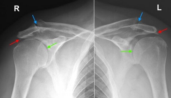
- Radiography of the shoulder joint.Dystrophic changes and growth of marginal bone (osteophytes) are found, at a later stage narrowing of the joint space, deformation and changes in the underlying bone structure are determined. Joint gaps can acquire a wedge -shaped shape, osteosclerotic changes and cyst formation can be seen on the bone.
- Tomographic research.In doubtful cases, especially in the early stages of the disease, CT of the shoulder joint is performed to obtain additional data on the condition of the bones and cartilage. If necessary to assess the condition of the soft tissue, magnetic resonance imaging is performed.
Differential diagnosis
The differential diagnosis of arthrosis is made with gout, psoriatic, rheumatoid and reactive arthritis, as well as pyrophosphate arthropathy. With arthritis, blood tests show signs of inflammation; changes on radiography are not very noticeable, osteophytes are absent, there are no signs of articular surface deformation.
In psoriatic arthritis, along with articular manifestations, skin rashes are often found. In rheumatoid arthritis, a positive rheumatoid factor is determined. With pyrophosphate arthropathy and gouty arthritis, biochemical blood tests show appropriate changes (increased levels of uric acid salts, etc. ).
Treatment of shoulder arthrosis
The patient is under the supervision of an orthopedic surgeon. You need to limit the load on the arms, excluding sudden movements, lifting and carrying prolonged loads. At the same time, it should be remembered that inactivity also has a negative effect on diseased joints. To keep the muscles in a normal state, as well as to restore the shoulder joint, you need to regularly perform a complex of exercise therapy recommended by a doctor.
Conservative treatment
One of the most pressing tasks in arthrosis is to fight the pain. To relieve pain and reduce inflammation, the following are prescribed:
- General action drugs.NSAIDs are prescribed in tablets during exacerbations. With uncontrolled use, they can irritate the stomach wall, negatively affecting the state of the liver and metabolism in cartilage tissue, therefore, they are taken only as directed by a doctor.
- Local medicine.NSAIDs are usually used in the form of gels and ointments. Self -administration may occur if symptoms arise or increase. More rarely, topical hormone preparations are indicated, which should be used in accordance with the doctor's recommendations.
- Hormones for intra-articular administration.In case of severe pain syndrome, which cannot be eliminated by other methods, intra-articular drug administration (triamcinolone, hydrocortisone, etc. ) is performed. Restrictions are imposed no more than 4 times a year.
To restore and strengthen cartilage in stage 1 and 2 arthrosis, agents from the group of chondroprotectors are used - drugs containing hyaluronic acid, chondroitin sulfate and glucosamine. The course of treatment is long (from 6 months to a year or more), the effect becomes noticeable after 3 months or more.
Physiotherapy treatment
With arthrosis of the shoulder joint, massage, physiotherapy exercises and physiotherapy techniques are actively used. During the remission period, patients are referred for spa treatments. Applying:
- mud and paraffin therapy;
- medicine bath;
- magnetic therapy and infrared laser therapy;
- ultrasound.
Surgery
In stage 3 of the disease, with significant cartilage destruction, limitation of movement and deformity, joint replacement is performed. References for surgery are given taking into account the age of the patient, his level of activity, the presence of severe chronic diseases. The use of modern ceramic, plastic and metal endoprostheses allows you to fully restore joint function. The lifespan of a prosthesis is 15 years or more.
Prediction
Arthrosis is a long -term and progressive disease. It is not completely cured, however, it is possible to slow the development of pathological changes in the joints, to maintain a high working ability and quality of life. To achieve maximum effect, the patient must seriously think about his illness and his willingness to follow the doctor's recommendations, even during the remission period.
Prophylaxis
Preventive measures include reducing household injuries, observing safety at work, eliminating excessive strain on the shoulder joint while performing professional duties and playing sports. It is necessary to diagnose and treat pathologies in a timely manner that can provoke the development of arthritis changes.